Layout/Characteristics
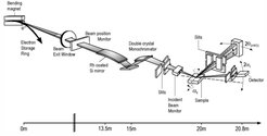
The beamline can either be operated in monochromatic, pink or white beam mode. The key parameters of the beamline are summarized in the table at the end of this section. The main optical elements are a Rh coated Si mirror and the double crystal monochromator (DCM). The mirror allows to cut the energy spectrum of the incident photons at its higher end to suppress the harmonic content in the monochromatic beam. In addition, it is used to focus the beam in the vertical direction. The DCM consists of a flat Si(111) single crystal and a sagittal Si(111) crystal bender for horizontal focusing. The position of the incident X-ray beam is traced by a blade beam position monitor in front of the optics. The outgoing beam can be monitored by the insertion of a fluorescence screen at the end of the optics. In case of the point detector, two pairs of horizontal and vertical slits allow to pre-select the beam size on the sample.

|
Source |
1.5 T Bending magnet (Ec = 6 keV), 0.3 mrad horizontal, 0.03 mrad vertical |
|
Energy range |
6 keV - 20 keV |
|
Optics |
Double crystal monochromator with a pair of Si(111) crystals, second crystal allows horizontal focusing of the beam |
|
Energy resolution (ΔE/E) |
3·10-4 @ 9 keV |
|
Flux at sample position |
1.0·10+12 ph/s/0.1% bw @ 9 keV |
|
Sample environment |
HT oven, cryostat, Kapton dome with He atmosphere, fast capillary spinner |
|
Beam size at sample |
could be focused to 0.3 mm (Hor) x 0.2 mm (Ver) |
|
Experimental setup / sample positioning |
Multiple circle diffractometer, horizontal / vertical sample normal geometry, horizontal / vertical four circle geometry, COR 5m from DCM |
|
Experimental setup / detectors |
Mythen 1K 1D-detector (50 μm x 8 mm, 700 Hz), |
|
Software: Control system / Data treatment / evaluation |
SPEC, software for reciprocal space mapping, software for reflectivity and crystal truncation rod analysis |
